
Early Pregnancy Scan – The Best Way to Detect Fetal Abnormalities
 An ultrasound scan is an absolute necessity and is part of the routine examination in any pregnancy. In a normal pregnancy, three scans are usually done; the first one between 6 to 9 weeks, the second between 11 to 14 weeks and the last between 18 to 23 weeks. The first two falls under the category of early pregnancy scans and are greatly helpful for the gynecologist to determine if the pregnancy as well as the fetus is developing normally.
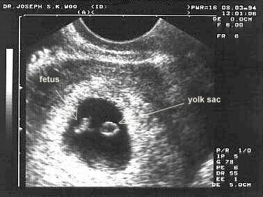
The early pregnancy scan which is done during 6 to 9 weeks is also known as dating or viability scans. The gynecologist carries out this ultrasound scan to confirm the location of pregnancy; this one can pick up ectopic pregnancies where the fetus grows in the fallopian tubes instead of the uterus. This scan can also be used to identify the number of fetuses as in a singleton or multiple pregnancies and also confirm and monitor fetal heart activity which is very vital. The doctor also uses this ultrasound to find out the age of the fetus and thereby provide a due date to the expectant mother.
The second ultrasonography is carried out during 11 to 14 weeks of pregnancy. Your obstetrician will be able to show you the fully formed physical image of your baby during this time. The doctor also makes use of this test to find out if there are any physical abnormalities in the fetus like missing limbs, extra fingers or any other deformity. This scan is also very vital for detecting genetic conditions like Downs syndrome.
The scan for Downss syndrome is also known as nuchal scan and is carried out during the 11th to 14th week of gestation. This scan is specifically recommended for all women who fall in the high risk category but can also be done in a routine pregnancy to avoid any nasty surprises after delivery. Many women opt for a nuchal scan as it is the best non invasive method to detect Downs syndrome when compared to other options like chorionic villus sampling test which is not only invasive but also risky for the fetus.
In this scan, the nuchal translucency is measured which is basically the amount of fluid that is situated at the neck region of the baby. Nil or very little fluid is present in normal fetuses by the 14th week of gestation but in babies with Downs syndrome, the level of this fluid is much higher. The crown to rump length of the baby is also measured and the two are compared to find out if your baby could be suffering from this chromosomal defect.
The nuchal scan is not a confirmatory test and has to be done in conjunction with other tests to confirm Downs syndrome. But it is a very good indicator of this condition and hence is one of the most recommended early pregnancy scans. It is always important to consult a trained and skilled radiologist for your pregnancy scans as this test provides the maximum information about your baby during pregnancy.

